
Ensis minor (Chenu, 1843) and Ensis siliqua (Linnaeus, 1758) are often difficult to distinguish and according to mitochondrial DNA evidence (Vierna et al., 2008) the two species are closely related. Further to my description of the two species (Dansey, 1998), I offer a key in this article to determine the differences. I am indebted to von Cosel (2009) for additional information on the shell exterior.
Identification of the two species
The data for identification and distribution of the two species are from my own records and examination of British Museum collections. Illustrations can also be found on the Conchological Society website:- http://www.conchsoc.org/encyclopedia
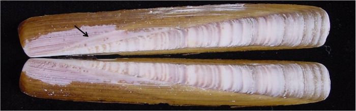
Ensis minor ( figure 1)
i. Slight groove on the outside of shell from the umbo region (i.e. the anterior/dorsal corner) to the middle of the ventral edge, sometimes just a change to lighter colouration (see arrow in figure).
ii. Articulated shells have a V-shape formed by the two valves.
iii. Holding the shell horizontally and looking from the ventral edge towards the dorsal edge the last third of the posterior end slopes down towards the posterior edge, so that looking at the posterior edge the two valves form a compressed gape like a Tudor arch.
iv. The growth ridges towards the posterior edge are close together.
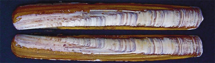
Ensis siliqua ( figure 2)
i. There is no groove.
ii. Articulated shells have two valves that curve into towards each forming a compressed O-shape.
iii. Holding the shell horizontally and looking from the ventral edge towards the dorsal edge the last third of the posterior end hardly slopes down towards the posterior edge, so that looking at the posterior end the two valves form an oval gape.
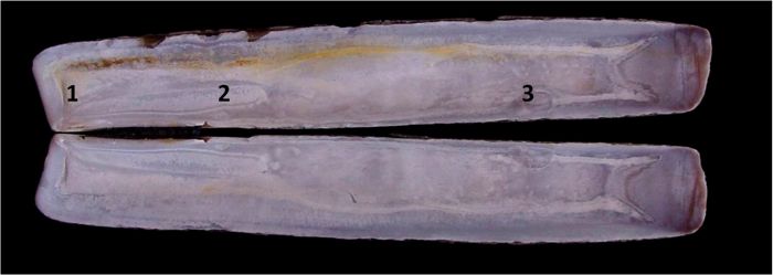
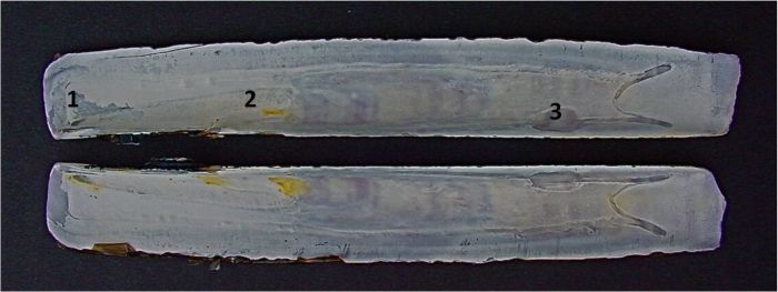
The following key applies to the interior shell muscle scars:
| Ensis minor – figure 3 | |
| Ensis siliqua – figure 4 | |
| 1. Anterior pallial scar | a) parallel to anterior edge b) much nearer to anterior edge than ventral pallial scar is to ventral edge |
| a) diverging from anterior end b) nearer to anterior edge than ventral pallial scar is to ventral edge | |
| 2. Anterior adductor scar | not much broadened posteriorly |
| broadens posteriorly | |
| 3. Posterior adductor scar | at own length or more from pallial sinus |
| at less than its own length from pallial sinus |
Data collection
My observations were made from the following stretches of coastline:
Ensis minor
North Norfolk Coast: from Hunstanton to Wells.
North Yorkshire: Saltburn.
Wales: Shell Island in Cardigan Bay.
Ensis siliqua
Lancashire: The Ribble Estuary.
North Wales: Prestatyn and Rhyl.
South Wales: Kidwelly, Pendine, Saudersfoot, Tenby and Oxwich Bay (The Gower Peninsula).
Ensis siliqua and Ensis minor
Scotland: Firth of Forth, St Andrews, Dumfries & Galloway.
South West England: South Cornwall, North and South
Devon, Dorset.
References
Cosel, R von (2009) The razor shells of the eastern Atlantic, part 2. Pharidae II: the genus Ensis Schumacher, 1817 (Bivalvia, Solenoidea). Basteria, 73: 9-56.
Dansey P (1998) A Proposed Key to the Identification of the Razor Shell Genus Ensis found in the British Isles. The Conchologists’ Newsletter 9(1) No. 145: 1 – 11.
Vierna J, Martinez-Lage A & Gonzalez-Tizon A M (2008) Molecular phylogeny of seven Ensis species based on mitochondrial DNA sequences, presented at the 5th Congress of the European Malacological Societies, Azores, Portugal.