|
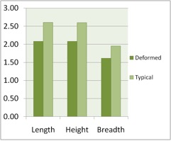
Recently I attended the field meeting described in the previous article, which included the shore of the Thames. The river at this is point muddy, tidal, mildly saline and fairly polluted, supporting a rather small number of extant mollusc species (and not much else). The introduced Corbiculid Corbicula fluminea is very common in this area (just upstream of Kew Railway Bridge), living in high concentrations on the mud surfaces exposed at low tide. Adrian Rundle, who was leading the meeting, drew my attention to an abnormal form present among the normal Corbicula. This form is rather unusual so I will describe it here as it may be of interest to members:- The shells of the aberrant form are generally small, rarely more than 25 mm in length, and rather globose in cross-section (as opposed to normal specimens, which tend to have a more heart-shaped cross-section). The posterior margin is generally more rounded than that of a typical example, and the ventral margins are turned inwards toward each other and often substantially deformed, leading to irregularities in the shape of the whole shell. The growth lines towards the shells’ margins are crowded and usually irregular. Internally, the growth margins are built up with a very distinctive, greenish or greyish callus*, usually extending from the pallial line to the margin itself, which sometimes spreads to the hinge plate rendering the teeth unrecognisable. According to Spann, Harper, and Aldridge (2010), the callus is made up of a form of calcium carbonate (vaterite) not usually found in the structure of Corbiculid shells (which are typically aragonitic) [see also note from Adrian Rundle below – Ed.]. A close examination of the shells shows that the callus is only laid down after a period of normal growth - the shell grows to a size of 10 to 20 mm in a normal fashion before the margins begin to turn inwards and the first layers of callus are laid down (once this begins to happen the shell does not increase appreciably in size, probably because growth is concentrated in the deformation structures). This is interesting as it points to a pathological origin of the abnormalities, not an environmental cause, as this would clearly affect the mollusc throughout its life and not just at a certain point in the growth sequence. Now for some statistics: at the Kew Bridge site, I collected a total of 198 specimens of dead C. fluminea, of which 68 showed some degree of callosity – 34% of the sample, or one in 2.91 specimens showing the abnormality. Deformed specimens from the site had an average width (anterior to posterior) of 20.8 mm, a height (top of umbone to lowest point on ventral edge) of 20.8 mm, and a breadth (side to side, both valves) of 16.1 mm. By comparison, typical, adult** specimens of C. fluminea taken in the sample had an average width of 26 mm, a height of 26 mm, and a breadth of 19.5 mm, making an average, normal specimen exactly 20% larger than the average deformed specimen (this is hardly surprising, given that, in abnormal shells, growth in terms of dimensions appears to slow down once the deformities begin to develop). More remarkable is the percentage of the specimens affected within my sample (provided it is taken as representative of this population, which it may not be) – the fact that the majority of specimens displayed no deformities seems to rule out purely ‘ecotypic’ variation (variation as a result of environmental factors), at the very least making it unlikely. Also, remarkably few specimens were found showing characteristics intermediate between the typical and abnormal forms. When presented with this one must be careful not to jump to conclusions – all that can be said is that deformation in C. fluminea is not caused solely by environmental factors (if this was the case, the entire population would display it to some degree). In view of this, it seems likely that the deformity of the shell margins is pathological in nature and is probably caused by a pathogen, perhaps a protozoan, which affects the molluscs’ growth-processes. It could potentially be caused by some genetic abnormality, but this seems less likely as if this was the case either the majority of my sample or a very small proportion would probably have been affected. In the same way we can rule out purely ecotypic variation, although environmental factors may contribute in some way. C. fluminea was first recorded in the UK in 1998. At present it is found in the River Thames and throughout the Broads system, and also on the River Barrow in Ireland. Similarly deformed specimens have been found in the Rivers Yare, Waveney, Thames and New Bedford, the different populations being affected to various degrees, those in the Yare showing deformities throughout (Spann et. al., 2010). I for one cannot find an explanation of these deformities in the literature, although they have been studied fairly extensively. If anyone can explain them, perhaps they could contact Mollusc World to make the matter clear.
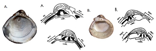
figure 2: Internal structure of typical and aberrant C. fluminea A. – Typical left valve, diagram of typical tooth structure B. – Left valve of aberrant spec., diagram of tooth structure in severely deformed spec. Abbreviations used:- ANT: antero-lateral teeth, POST: posterior lateral teeth, CARD: cardinal teeth. (Locality data as for Fig. 1).
*very large, adult specimens of the typical form often show a degree of callosity along the ventral margin. However this is distinct from the callus I describe, being generally dark slatey-grey in colour and running only along the shell edge, never extending up to the pallial line as in the deformed specimens. It results from the slowing-down of growth and can be seen in many bivalve species. **not many juvenile specimens were found. All those in the sample below two centimetres in length (only 14 specimens in total) were ignored when calculating the averages, as they were clearly juvenile and had the potential to develop into examples of either form.
References Killeen, I., Aldridge, D.C., & Oliver, G. (2004) Freshwater Bivalves of Britain and Ireland. FSC (contains general description of C. fluminea) Spann, N., Harper, E.M. & Aldridge D.C. (2010) The unusual mineral vaterite in shells of the freshwater bivalve Corbicula fluminea from the UK. Naturwissenschaften, 97, 743-751.- (chemical makeup of deformation structures in C. fluminea) http://www.zoo.cam.ac.uk/zoostaff/aldridge/nicole.htm - N. Spann (freshwater mussels as biomonitors) http://caisie.ie/?p=233 (Corbicula fluminea in the River Barrow, Ireland) |
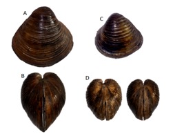
figure 1: Typical and abnormal specimens of Corbicula fluminea A. – Typical specimen, right valve B. – Typical spec., view of anterior C. - Abnormal spec., left valve D. – Abnormal spec., view of anteriors (all specimens collected under Kew rail Bridge (TQ[51]195775), Kew, London, 9.7.2011).
figure 3: Graph showing length, height and breadth of typical and aberrant specimens in my sample (all values represent centimetres). |
Pathological causes for deformation in Corbicula fluminea?
Issue
27
Page
8