|
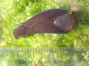
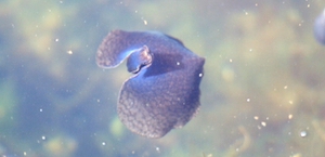
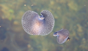
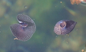
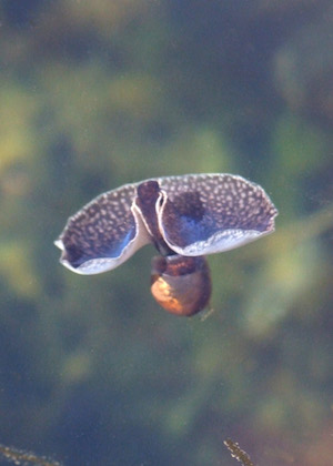
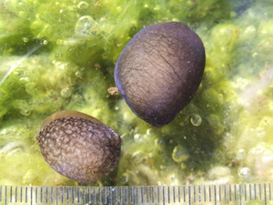
Late in March 2010, as I was walking past a small seawater pond near Oban in west Scotland, I caught sight of an extraordinary animal swimming with great energy and panache. Others appeared alongside it and together, just below the water surface, they performed an unforgettable display of synchronised swimming. They were rather like bats flying slowly underwater, except that each had a near circular membrane as its “wings”. I had never seen anything like it. I soon identified the animal as Akera bullata, an opisthobranch gastropod. This article describes some of the findings that arose out of that discovery. One of the most intriguing aspects of this marine mollusc is the variety of bodily shapes and postures that it can adopt. These are so diverse that it can be hard to believe they all involve the same animal. For most of the year Akera is a dark, drab sea-snail. When small, it crawls on and eats seaweed; when large, it prefers silty sediment. Its most notable features are the rectangular head, the two parapodia (lateral extensions of the foot) that fold over its back, and the thin, delicate shell partly visible at its rear end (figure 1). It often moves and feeds under silty debris and can sometimes be hard to see. For a short period in the spring, just before mating and spawning, Akera is almost magically transformed. Shoals of swimmers perform an elegant underwater ballet. Their beauty is enhanced by a glancing blue iridescence, probably caused by interference of light in parts of the integument (figure 2). During the powerful downstroke of the “wings” (parapodia), the body surface becomes so tightly stretched by the strong swimming muscles that the yellow colour of the thin shell, and through that the brighter yellow of the digestive gland, are briefly visible (figure 3). The few scientists who have seen Akera swimming have abandoned their sober scientific style and, quite independently, waxed lyrical about its bizarre beauty:
“The movements have an extraordinary lightness and grace” (Morton and Holme, 1955).
“…no-one can possibly see it and forget it.” (Yonge and Thompson, 1976).
“The animal… hovers in the most graceful postures in the clear water…The movements are so charming that we wish we could make it swim whenever we wanted. However, although there are almost always Akera in our aquarium, we rarely see them swimming.” (Meyer and Möbius, 1865).
“Nothing is more interesting and graceful than Akera when it is swimming ..” (Guiart, 1901).
Guiart describes his journey on foot to see this spectacle at a remote site on the Atlantic coast of France. He not only risked his life in quicksands but, while wading up to his thighs, was briefly attacked by a 1.5 m long octopus “with arms as thick as your wrist”. But he urged his readers:
“What you are going to see will more than make up for these petty irritations, so just ignore them. ..… Where sun warms the deep water, you will see small, elegant animals rising from the sand and flying up to the surface. They sink and rise again, appearing from left and right, exultantly beating wings that undulate like the costume of a miniature Loie Fuller. You will be entranced by this new Serpentine Dance. ” [Loie Fuller (1862-1928) was a famous dancer who devised many original dances. One of her creations, the Serpentine Dance, involved huge, rapidly undulating, expanding and contracting circular veils on each arm. This dance does indeed bear some resemblances to Akera’s swimming style, as can be seen in a performance by Fuller filmed in 1896 and now on You Tube.]
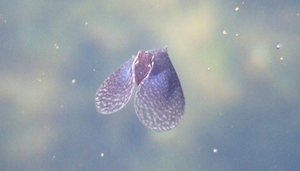
Akera swims by powerful synchronised beats of its two parapodia (Morton and Holme, 1955). These wing-like flaps are broadly united with the foot and, at the start of the downstroke, the three together form the swimming membrane, a near-complete medusa-like disc. The downstroke is rapid and powerful, driving the animal upwards and keeping it waterborne (figure 4). At the end of the downstroke, the wings are wrapped tightly around the body, making a streamlined shape with a sharply pointed rear end (figure 5). Immediately, the animal begins to sink under the weight of its shell and internal organs. It continues to sink during the much slower upstroke when, paradoxically, the relaxed edges of the wings are turned upwards because the animal is sinking faster than the wings are moving upwards (figure 6). [The diagrams given by Thompson (1976, figures 14a and 14b on page 29), repeated in Thompson & Brown (1976, figures 11a and 11b on page 30) and elsewhere, wrongly show this upward turning of the edges to be a feature of the downstroke, as common sense would suggest it to be. In fact, during the downstroke the muscles are so tightly contracted and the wings so rigid that the edges remain firm (figure 4).] Another author made a more fundamental, if understandable, error. Watching Akera swimming, it is easy to gain the false impression that the flapping cloak-like “wings” are attached to its back and that their outer edges meet along its ventral surface – perhaps because it looks rather like a cloaked human being. In fact the reverse is true – the “wings” (parapodia) are attached to the ventral foot and they meet over the animal’s back. This illusion was probably the cause of the impossible drawing in Clayton (1974, page 147) showing a swimming Akera with its ventral surface and shell mouth visible through the closing “wings”. In 2010 the number of individuals swimming in the pond at any one time exceeded 100 on two days (5 and 8 April) and was in the range 20-100 on five other days in April. This number declined sharply after mid-April and almost none were seen swimming during the rest of the year, indicating a brief but intense swimming period. There was a swimming season again in April 2011 but, for unknown reasons, the numbers swimming were much lower, rarely exceeding single figures at any one time. Why do Akera swim? It has been suggested that it is a form of sexual display, since it has usually been recorded shortly before spawning (Thompson, 1976). This cannot be the whole story, however. Akera of all sizes swim, except the really big ones (the largest swimmer I have seen weighed 6.5 g). In April 2010 I saw small numbers, up to twenty on each occasion, of minute, recently-metamorphosed Akera swimming together in the pond shallows. Still translucent and largely unpigmented, they swam with the same postures and movements as adults (figure 7). Of a sample of 13, one weighed only 18 mg and all were less than 80 mg. I have the impression that Akera swim to escape undesirable conditions, such as overcrowding, heat or food shortage. Akera adopts various other postures. Hedgehog-like, it rolls into a ball when handled or provoked (figure 8). It cannot withdraw completely into its shell and, in this position, the parapodia are wrapped tightly around the shell and the withdrawn head. During mating, Akera contorts and stretches its body (figure 9). It is a hermaphrodite and copulation is a communal activity performed in chains; each animal acts as male to the one in front and female to the one behind. Usually the chains are coiled in on themselves and hidden under debris, so it is difficult to see what is happening. I have encountered groups of up to six assembled in this way, presumably mating. The Oban Akera pond measures ca 25 x 5 x 1.5 m deep and held at least 2,800 individuals in spring 2010. This estimate was based on the number of empty shells and dead and dying animals at the pond edge during summer. Akera lives for about a year and dies soon after spawning, many or most crawling ashore to die. Earlier, I had intended to measure the number in the pond by a mark-and-recapture method. To find a suitable marking technique, I test-marked the shells of a few live specimens with a small dot of correcting fluid (Tippex). Sadly they all died within a few hours. Unlike prosobranchs and land snails, Akera have thin uncalcified shells – hence the name “bubbleshells” for this group of opisthobranchs. Solvent must have rapidly entered their bodies through the shell. How is it that Akera can reach such a high density in a small pond like this, but seem rarely to be met with in such numbers in the open sea or in firths and estuaries? Probably because the pond has no outlet. It is a settling pond and soakaway, dug about ten years ago to take the outflow from a marine aquarium. Akera larvae have a very brief pelagic stage and, presumably, a few larvae entered the pond in the water supply years ago and gave rise to this population. Moreover, there are no fishes or other predators such as large crabs in the pond. In such conditions, invertebrates rule! A similarly isolated population was recorded by Morton and Holmes (1955) in a seawater tank in Plymouth naval dockyard and was used in their classic photographic study. Another well-known population, in more natural conditions but not fully isolated from the open sea, occurs at the Fleet in Dorset (Thompson and Seaward, 1989). It is near-impossible to measure the body length of an animal that extends and contracts as it moves, and contracts strongly as soon as you approach it! With time and patience it can be done for one animal, but measuring individual lengths of large samples is wholly impractical. I therefore used live mass as a standard measure of size, weighing each after blotting it gently dry with absorbent tissue paper. The largest from the pond, where the substrate and main diet of larger Akera is a silt-sand mixture, weighed 11.0 g. However, two raised from the egg in captivity on a silt substrate attained 19.2 and 14.6 gm. Like others, they spawned repeatedly and lost much weight in the month before death. Their large size may have been attributable to the silt, or lower population density, or other conditions of captivity. The shell of the largest was 33 mm long, somewhat smaller than the 41 mm maximum recorded by Thompson and Seaward (1989) for specimens from Lough Ine in Ireland. Finally, something remarkable. One day, the largest captive-bred Akera escaped from the tank. It ended up on a filter, a perforated metal sheet at the drain where waste water leaves the aquarium. The holes in the metal sheet were six mm in diameter. The large Akera was about 10 mm wide at the head and 20 mm at the parapodia. Yet it had squeezed its foreparts, including the parapodia, through one of the holes and had been stopped only by its 23 mm wide shell. It couldn’t move in either direction and was firmly stuck. Because of the large width and bulk of body that had gone through, it was impossible to draw it backwards without killing it. Sadly, my prize specimen died, and a slightly smaller one met the same fate some days later. The extraordinary volume contraction that must have accompanied this feat presumably involves a large redistribution of body fluids, together with deformation of the soft integument. The same thing must happen when large terrestrial slugs escape from sprung mousetraps (the breakback variety), or enter houses by squeezing under closed, tightly-fitting modern doors – phenomena I suspect others will also have experienced?
Acknowledgements I am grateful to Gill Notman for her inspiration and enthusiasm in setting up the captive rearing tank, and to Helen Marshall for advice on the larval stage of Akera bullata. Clayton, M. (1974) The Living Seashore. Warne, London. Guiart, J. (1901) Contribution a l’étude des gastéropodes opisthobranches et en particulier des cephalaspides. Mémoires de la Société Zoologique de France, 14: 5-219. Meyer, H.A. and Möbius, K. (1865) Fauna der Kieler Bucht, Band. 1. Die Hinterkiemer oder Opisthobranchia. Engelmann, Leipzig. Morton, J.E. and Holme, N.A. (1955) The occurrence at Plymouth of the opisthobranch Akera bullata, with notes on its habits and relationships. Journal of the Marine Biological Association of the United Kingdom 34: 101-112. Thompson, T.E. (1976) Biology of Opisthobranch Molluscs Vol 1. The Ray Society, London. Thompson, T.E. and Brown, G.H. (1976) British Opisthobranch Molluscs. Synopses of the British Fauna (New Series) No 8. Linnaean Society, London. Thompson, T.E. and Seaward, D.R. (1989) Ecology and taxonomic status of the Aplysiomorph Akera bullata in the British Isles. Journal of Molluscan Studies 55: 489-496. Yonge, C.M. and Thompson, T.E. (1976) Living marine Molluscs. Collins, London |
Figure 1: A medium-sized Akera bullata, crawling. (Photo: Clive Craik)
Figure 2: Blue iridescence in a swimming Akera (Photo: Clive Craik)
Figure 3: Two Akera swimming. Left animal, heart-shape at start of downstroke. Right animal, side view, start of upstroke. (Photo: Clive Craik)
Figure 4: Wings are rigid during the rapid, powerful downstroke (Photo: Clive Craik)
Figure 5: Two Akera swimming. Left: near end of upstroke. Right: end of downstroke, tightly-wrapped parapodia and pointed rear end. (Photo: Clive Craik)
Figure 6: Mid-upstroke, dorsal view: edges are upturned because animal is sinking. (Photo: Clive Craik)
Figure 7: Two very small Akera (< 50 mg each) swimming. Left: end of downstroke, tightly-wrapped and pointed. Right: heart-shape of early downstroke. (Photo: Clive Craik)
Figure 8: Two Akera rolled up in defensive posture after being handled. (Photo: Clive Craik)
Figure 9: Mating assembly of three Akera. (Photo: Clive Craik)
|
The Many Faces of Akera bullata
Issue
28
Page
5
Species